Editor: Tiffany
Scientists have developed a new method using magnetic nanoparticles coated with glucose and Safranal to selectively target and kill liver cancer cells, offering a promising approach to reduce the side effects of traditional chemotherapy.
Key Highlights
- Research Question:
Can iron oxide nanoparticles coated with glucose and conjugated with Safranal effectively target and induce apoptosis in liver cancer cells? - Research Difficulties:
Overcoming the limitations of current liver cancer treatments, such as the non-specificity and side effects of chemotherapy, while ensuring the nanoparticles are biocompatible and stable. - Key Findings:
The nanoparticles demonstrated a significant antiproliferative effect on HepG2 cells with an IC₅₀ of 305 μg/mL, increased ROS levels, induced apoptosis, and caused cell cycle arrest at the G2/M phase. - Innovative Aspects:
The use of glucose coating to enhance uptake by cancer cells and the conjugation of Safranal, a natural compound, to magnetic nanoparticles for targeted delivery. - Importance of the Study:
This research provides a potential new strategy for liver cancer treatment that could improve efficacy and reduce side effects, paving the way for further in vivo studies and clinical trials.
The Urgent Need for Better Liver Cancer Treatments
Liver cancer ranks among the deadliest cancers worldwide, claiming over 830,000 lives annually and affecting more than 905,000 new patients each year, according to global health estimates. Known medically as hepatocellular carcinoma (HCC) when it originates in the liver, this disease often progresses silently in its early stages, with symptoms like fatigue, abdominal pain, jaundice, and unexplained weight loss emerging only as it advances. By then, treatment becomes far more challenging. For patients diagnosed with advanced or metastatic liver cancer, options like surgery may no longer be viable, leaving chemotherapy as a common recourse. However, chemotherapy’s broad attack on both cancerous and healthy cells often leads to debilitating side effects, such as nausea, hair loss, and weakened immunity, while its effectiveness remains limited against aggressive tumors. This lack of precision in current treatments highlights a critical need for therapies that can zero in on cancer cells without harming the rest of the body. Researchers have long sought innovative solutions to this problem, and a new study from Iran offers a promising step forward.
A Novel Nanoparticle Approach to Combat Liver Cancer
A team of scientists from the Islamic Azad University, Rasht Branch, Iran, set out to tackle liver cancer with a cutting-edge approach: magnetic nanoparticles designed to deliver drugs directly to tumor cells. Their goal was to create a system that could enhance the effectiveness of anticancer treatments while reducing the toxic fallout seen in conventional therapies like chemotherapy. Led by Somayeh Mikaeili Ghezeljeh, Ali Salehzadeh, and Somayeh Ataei-e Jaliseh, the researchers developed iron oxide nanoparticles coated with glucose and conjugated with Safranal—a natural compound from saffron with known anticancer properties. They aimed to test whether these nanoparticles could selectively target and kill liver cancer cells, specifically the HepG2 cell line, by triggering apoptosis, a process of programmed cell death. Their work, published in BMC Chemistry in 2024, sought to harness the magnetic properties of the nanoparticles to guide them to cancer sites using an external magnetic field, potentially revolutionizing drug delivery for liver cancer patients.
From Lab to Cell: Testing the Nanoparticles’ Power
The study unfolded in several meticulous steps, from crafting the nanoparticles to testing their effects on cancer and normal cells. Here’s how the researchers approached it and what they found:
1. Synthesis and Characterization of Nanoparticles
- Procedure: The team synthesized iron oxide nanoparticles (Fe₃O₄) using iron chloride salts and ammonia, then coated them with glucose to improve biocompatibility and uptake by cancer cells. Safranal was chemically attached to the glucose coating to complete the nanoparticles, dubbed Fe₃O₄@Glu-Safranal NPs.
- Result: Analysis using techniques like FT-IR, XRD, and TEM revealed spherical nanoparticles with sizes ranging from 17 to 49 nm, a surface charge of -13 mV, a hydrodynamic size of 129 nm, and magnetic saturation of 22.5 emu/g.
- Finding: The nanoparticles were pure, stable, and well-suited for medical use, with their magnetic properties intact for targeted delivery.
2. Antiproliferative Effect (MTT Assay)
- Procedure: Liver cancer cells (HepG2) and normal human cells (HEK293) were exposed to varying concentrations of the nanoparticles for 24 hours, and cell viability was measured using the MTT assay.
- Result: The 50% inhibitory concentration (IC₅₀) was 305 μg/mL for HepG2 cells and 680 μg/mL for HEK293 cells. By comparison, Safranal alone had an IC₅₀ of 474 μg/mL, while Cisplatin, a standard chemotherapy drug, had an IC₅₀ of 135 μg/mL.
- Finding: The nanoparticles were more toxic to cancer cells than normal cells, indicating a selective effect that could minimize harm to healthy tissue—a stark improvement over traditional drugs.
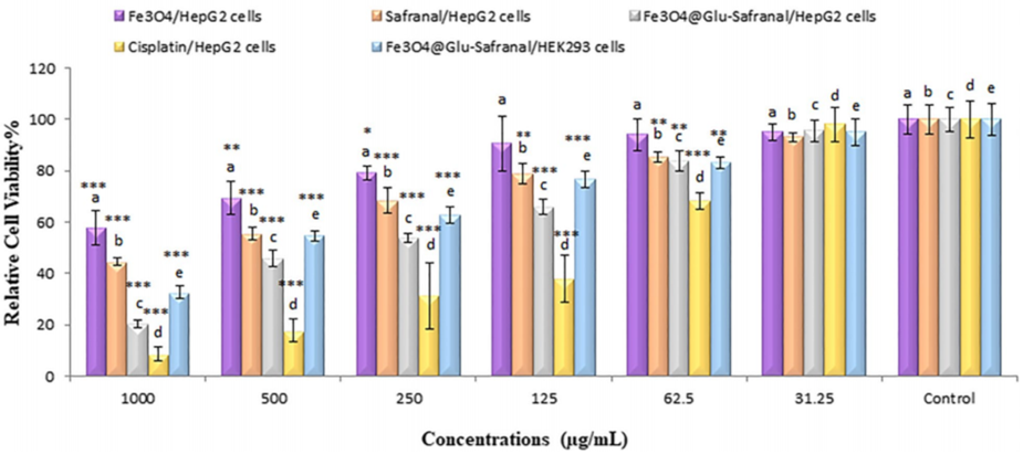
Figure 1. MTT assay results displaying the IC₅₀ values of Safranal, Fe₃O₄, Fe₃O₄@Glu-Safranal, and Cisplatin on liver cancer cells.
3. ROS Level Measurement
- Procedure: HepG2 cells were treated with the nanoparticles, and reactive oxygen species (ROS) levels were measured using a fluorescence assay to assess oxidative stress.
- Result: The percentage of cells with elevated ROS (DCFH+) rose from 1.97% in untreated cells to 19.60% in treated cells.
- Finding: The nanoparticles triggered a significant increase in ROS, likely damaging cancer cells by overwhelming their defenses, a key mechanism in their anticancer action.
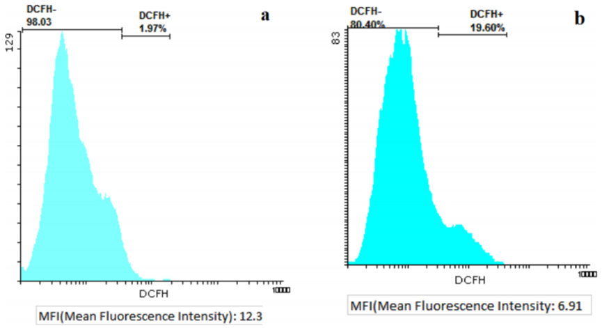
Figure 2. Flow cytometry analysis indicating increased ROS levels in HepG2 cells treated with Fe₃O₄@Glu-Safranal nanoparticles.
4. Cell Apoptosis/Necrosis Analysis
- Procedure: Flow cytometry quantified the extent of apoptosis and necrosis in HepG2 cells after nanoparticle treatment.
- Result: Apoptotic cells increased from 2.5% in untreated samples to 34.7% in treated ones, while necrosis rose to 4.88%.
- Finding: The nanoparticles predominantly induced apoptosis—controlled cell death—rather than messy necrosis, which is ideal for safe and effective cancer treatment.
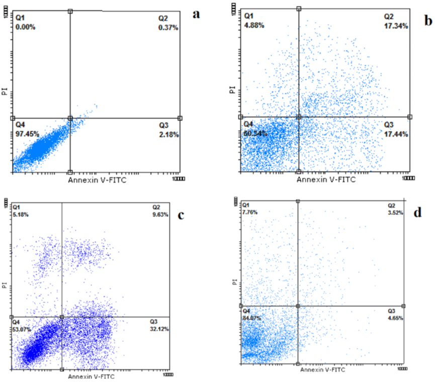
Figure 3. Flow cytometry scatter plots demonstrating the induction of apoptosis in HepG2 cells by Fe₃O₄@Glu-Safranal nanoparticles.
5. Cell Cycle Analysis
- Procedure: Flow cytometry examined how the nanoparticles affected the cell cycle phases in HepG2 cells.
- Result: The proportion of cells in the G2/M phase jumped from 13.9% in controls to 18.3% in treated cells.
- Finding: By halting the cell cycle at G2/M, the nanoparticles prevented cancer cells from dividing, further curbing tumor growth.
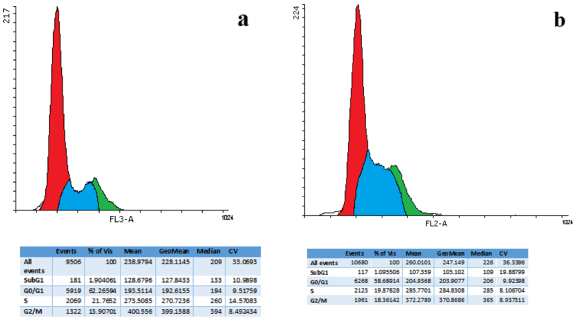
Figure 4. Cell cycle distribution histograms showing G2/M phase arrest in HepG2 cells treated with Fe₃O₄@Glu-Safranal nanoparticles.
Together, these experiments showed that Fe₃O₄@Glu-Safranal NPs not only targeted liver cancer cells effectively but also disrupted their survival mechanisms through multiple pathways—oxidative stress, apoptosis, and cell cycle arrest.
A Step Closer to Precision Medicine for Liver Cancer
This study unveils a potential game-changer in the fight against liver cancer. The Fe₃O₄@Glu-Safranal nanoparticles demonstrated a remarkable ability to home in on HepG2 cancer cells, killing them with precision while largely sparing normal cells. The use of glucose coating enhances uptake by cancer cells, which often crave sugar, while the magnetic properties allow for directed delivery—an elegant solution to chemotherapy’s scattershot approach. Safranal, already prized for its anticancer potential, gains new potency when paired with this nanotechnology. With an IC₅₀ of 305 μg/mL against cancer cells compared to 680 μg/mL for normal cells, the nanoparticles offer a safer, more targeted alternative that could reduce the harsh side effects patients endure.
Reference:
Mikaeili Ghezeljeh, Somayeh, Ali Salehzadeh, and Somayeh Ataei-e Jaliseh. “Iron oxide nanoparticles coated with Glucose and conjugated with Safranal (Fe3O4@ Glu-Safranal NPs) inducing apoptosis in liver cancer cell line (HepG2).” BMC chemistry 18.1 (2024): 33.