Editor: Tiffany
Researchers have developed a multifunctional nanoparticle system that combines quantum dots, silica, and gold nanoparticles to deliver targeted chemotherapy, enhance radiotherapy, and provide multimodal imaging for colorectal cancer, demonstrating improved treatment efficacy in preclinical models.
Key Highlights
- Research Question:
How can nanotechnology be utilized to create a targeted system that simultaneously delivers chemotherapy, enhances radiotherapy, and enables multimodal imaging for colorectal cancer? - Research Difficulties:
Overcoming limitations of current colorectal cancer treatments, such as non-specific drug delivery, insufficient tumor imaging, and resistance to therapy, while ensuring the nanoparticle system is biocompatible and stable in vivo. - Key Findings:
The hybrid nanoparticle system demonstrated selective targeting of colorectal cancer cells, improved drug delivery, enhanced radiotherapy efficacy, and clear tumor visualization through fluorescence, MRI, and CT imaging in both in vitro and in vivo models. - Innovative Aspects:
The integration of quantum dots for fluorescence imaging, mesoporous silica for drug loading, gold nanoparticles for radiotherapy enhancement and CT imaging, and EpCAM aptamers for targeted delivery into a single, multifunctional nanocarrier. - Importance of the Study:
This research presents a potential advancement in colorectal cancer theranostics by offering a targeted, multimodal approach that could improve treatment precision, reduce side effects, and enable real-time monitoring of therapeutic outcomes.
Challenges in Colorectal Cancer Treatment and the Role of Nanotechnology
Colorectal cancer (CRC) ranks as the third most common cancer globally, affecting millions and posing a major public health challenge. This malignancy, which develops in the colon or rectum, often becomes resistant to treatment and has a high recurrence rate, complicating patient outcomes. Symptoms such as persistent changes in bowel habits, blood in the stool, abdominal pain, and unexplained weight loss can signal its presence, but these often appear late, delaying diagnosis. Traditionally, doctors rely on colonoscopy—a procedure that, while effective, is invasive and uncomfortable for patients—to detect CRC.
Current treatment options include surgery to excise tumors, chemotherapy to attack cancer cells, and radiotherapy to shrink or destroy malignant tissue. However, these approaches have notable limitations. Surgery may leave behind microscopic cancer cells, chemotherapy affects healthy tissues alongside cancerous ones, causing side effects such as nausea and fatigue, and radiotherapy faces challenges in targeting tumors precisely. These limitations highlight the need for alternative approaches.
Nanotechnology is advancing cancer care by utilizing small particles to deliver drugs and imaging agents directly to tumors. Quantum dots (QDs) are semiconductor particles that fluoresce under light, aiding in tumor visualization. Mesoporous silica nanoparticles (MSNs) serve as drug carriers due to their porous structure, while gold nanoparticles (Au NPs) enhance radiotherapy and improve imaging contrast. These nanomaterials collectively provide a more targeted approach to managing CRC.
Developing a Multifunctional Nanoparticle System for Targeted Theranostics
The researchers aimed to develop a multifunctional nanoparticle system for targeted chemotherapy, enhanced radiotherapy, and tumor imaging in CRC. Specifically, they sought to synthesize a nanocarrier integrating quantum dots, silica, and gold nanoparticles, evaluate its performance in laboratory and animal models, and assess its imaging capabilities.
The study was conducted by Maryam M. Matin from Ferdowsi University of Mashhad, Iran, and published in Communications Biology in 2024.
Synthesis, Characterization, and Preclinical Evaluation of the Hybrid Nanocarrier
The researchers constructed a hybrid nanoparticle system through a stepwise process. They started with quantum dots (GZCIS/ZnS), valued for their fluorescence and low toxicity, and coated them with mesoporous silica to form QD@MSN. The pores of these particles were loaded with epirubicin (EPI), a chemotherapy drug. Gold nanoparticles capped the pores to retain the drug until it reached the tumor site. The system was PEGylated—coated with polyethylene glycol—for stability in biological environments and conjugated with EpCAM aptamers to target CRC cells specifically.
Key Experiments
- Synthesis and Characterization
- Procedure: Quantum dots were synthesized and coated with silica using a modified Stöber method, followed by EPI loading and capping with Au NPs. PEGylation and aptamer conjugation completed the system.
- Result: The nanoparticles had an approximate diameter of 65 nm and exhibited high drug-loading efficiency.
- Finding: The hybrid system was successfully synthesized with the intended size and drug-carrying capacity.
- In Vitro Studies
- Procedure: The nanoparticles were tested on CRC cells (HT-29) and normal cells (CHO) using assays such as MTT, flow cytometry, and confocal microscopy.
- Result: Targeted nanoparticles demonstrated higher uptake and cytotoxicity in HT-29 cells compared to non-targeted nanoparticles or free EPI.
- Finding: The system selectively targets CRC cells, improving drug delivery and cancer cell death.
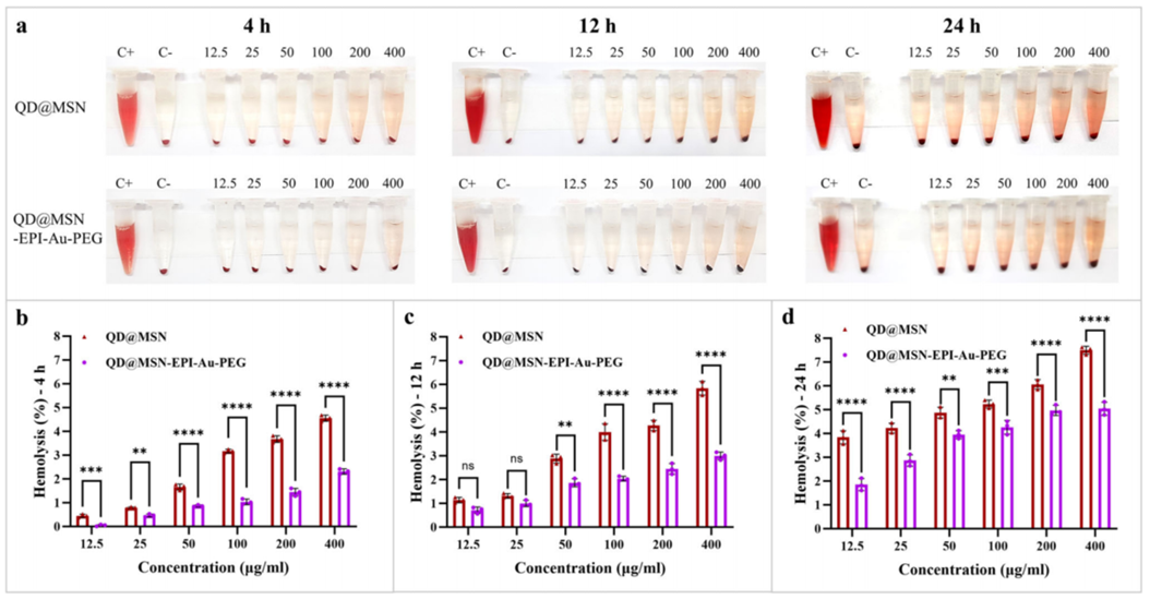
Figure 1. Hemolysis assay on backbone and PEGylated nanoparticles at three time points.
- In Vivo Therapeutic Efficacy
- Procedure: Mice with HT-29 tumors were treated with the nanoparticles and radiotherapy, with tumor size and survival monitored over time.
- Result: The combination treatment reduced tumor growth and increased survival compared to control groups.
- Finding: The hybrid nanoparticles enhance the efficacy of chemo-radiotherapy in animal models.
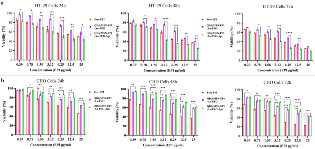
Figure 2. Tumor volume reduction (a) and survival curves (b) in treated mice.
- Imaging Studies
- Procedure: Fluorescence imaging, MRI, and CT scans were used to track nanoparticle distribution in mice.
- Result: The nanoparticles accumulated in tumors, generating clear signals across all imaging modalities.
- Finding: The system supports multimodal imaging, aiding in tumor detection and treatment monitoring.
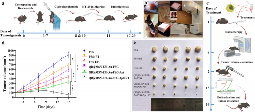
Figure 3. Fluorescence (a), MRI (b), and CT © imaging showing tumor accumulation.
Implications of the Nanoparticle System for Future Colorectal Cancer Management
This study introduces a novel hybrid nanoparticle system for colorectal cancer. By combining quantum dots, silica, and gold nanoparticles, it delivers chemotherapy to cancer cells, enhances radiotherapy effects, and enables detailed tumor imaging. The system showed improved targeting and efficacy in preclinical models, indicating potential for reduced side effects.
The multifunctional design allows for simultaneous therapy and imaging, which may facilitate treatment monitoring. The study notes limitations, such as the need for further optimization and safety assessments. Future efforts will focus on clinical translation and exploring applications in other cancers.
Reference:
Abrishami, Amir, et al. “Hybridized quantum dot, silica, and gold nanoparticles for targeted chemo-radiotherapy in colorectal cancer theranostics.” Communications Biology 7.1 (2024): 393.