Editor: Nina
Scientists develop biodegradable polyphosphoester micelles that function as dual agents for background-free magnetic resonance imaging and effective drug delivery in cancer treatment.
Key Preview
- Research Question: The study investigates the potential of biodegradable polyphosphoester micelles as dual-function agents for background-free magnetic resonance imaging (MRI) and drug delivery.
- Research Design and Strategy: The authors designed and synthesized polyphosphoester micelles, optimizing their structure for enhanced MRI properties and testing their ability to act as drug carriers.
- Method: The research involved the creation of amphiphilic gradient-type copolymer micelles, followed by in vitro and in vivo assessments of their imaging capabilities and drug delivery efficacy.
- Key Results: The polyphosphoester micelles demonstrated effective imaging in vivo without background interference and facilitated successful drug delivery of the therapeutic agent PROTAC.
- Significance of the Research: This work presents a sustainable alternative to traditional MRI agents and offers a novel approach to theranostic applications in medicine.
Introduction
In recent years, cancer has emerged as one of the leading causes of morbidity and mortality worldwide, characterized by the uncontrolled growth and spread of abnormal cells. Traditional cancer treatments, including chemotherapy and radiation therapy, aim to target and eliminate cancerous cells. However, these therapies often come with significant side effects, as they can also affect healthy cells, leading to a range of complications such as nausea, fatigue, and increased susceptibility to infections.
A common strategy for delivering drugs in traditional cancer treatments involves the use of systemic chemotherapy, where medications are administered intravenously or orally to target rapidly dividing cancer cells. While this approach can be effective, it presents distinct challenges, including poor bioavailability, limited drug distribution to tumor sites, and the potential for systemic toxicity. These challenges can result in inadequate therapeutic responses, resistance to treatment, and harm to the patient’s overall health.
To address these limitations, innovative drug delivery strategies have been developed, focusing on improving the efficacy and safety of cancer therapies. One such strategy is the use of nanocarriers, which can encapsulate therapeutic agents and selectively deliver them to tumor sites while minimizing exposure to healthy tissues. By utilizing biodegradable polymers as nanocarriers, researchers aim to enhance the targeting capabilities of drugs, reduce side effects, and improve patient outcomes. This study introduces biodegradable polyphosphoester micelles as a novel approach for drug delivery and imaging, showcasing their potential to serve as dual-function agents that can provide effective cancer treatment while facilitating real-time monitoring of therapeutic distribution through magnetic resonance imaging (MRI).
Research Team and Aim
The research was led by Olga Koshkina, who conducted this study during her tenure at the University of Twente. The multidisciplinary team included contributions from Timo Rheinberger, Vera Flocke, and several other collaborators from esteemed institutions such as Heinrich Heine University and the Fraunhofer Institute. Their paper, titled “Biodegradable polyphosphoester micelles act as both background-free 31P magnetic resonance imaging agents and drug nanocarriers,” was published in the journal Nature Communications.
The primary aim of the research, as articulated by the lead researcher, was to “develop a new class of biodegradable polymers that can serve both as MRI agents and drug carriers, paving the way for sustainable biomedical applications.” This innovative approach seeks to address the limitations of conventional imaging agents and drug delivery systems while promoting environmentally friendly solutions in medicine.
Experimental Process
Primary Technique
The primary technique employed in this study was the synthesis and characterization of biodegradable polyphosphoester micelles, which serve as dual-function agents for background-free magnetic resonance imaging (MRI) and drug delivery. This approach combined polymer chemistry with advanced imaging techniques to achieve the research objectives.
Experiment 1: Synthesis of Micelles
Key Steps:
- Preparation of Monomers: The researchers synthesized amphiphilic gradient-type copolymers using anionic ring-opening polymerization of polyphosphoesters containing phenyl and ethyl functional groups.
- Self-Assembly: The resulting copolymers were dispersed in aqueous solutions, allowing them to self-assemble into micelles with a defined core-shell structure.
- Characterization: Dynamic light scattering (DLS) was performed to determine the size and polydispersity index (PDI) of the micelles, ensuring they were within the desired range for optimal imaging and drug delivery.
Data Collection and Analysis:
DLS measurements were conducted at a scattering angle of 90° and at 295 K. The data were analyzed using ZSxplorer software to derive the average hydrodynamic radius and PDI of the micelles.
Result:
The synthesized micelles exhibited a hydrodynamic radius of approximately 9 nm with a PDI of less than 0.1, indicating a uniform size distribution suitable for biological applications.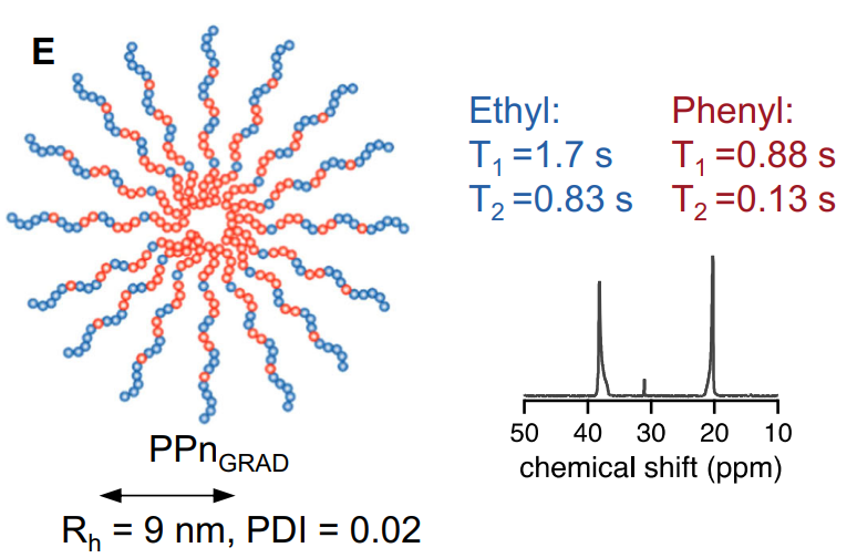
Figure 1. Schematic of PPnGRAD micelles with NMR spectrum (158 MHz, D2O).
Novel Aspects:
This experiment introduced a gradient microstructure to the micelles, which improved MRI characteristics by enhancing the mobility of the polymer chains, leading to longer T2 relaxation times compared to traditional block copolymer systems.
Experiment 2: In Vitro MRI Characterization
Key Steps:
- Preparation of Samples: Aqueous dispersions of the synthesized micelles were prepared at various concentrations (4 wt% polymer) for MRI analysis.
- MRI Scanning: The samples underwent 31P MRI using a spin-echo sequence on a 9.4 T MRI scanner with a fixed field of view (FOV) and predefined matrix size.
- Optimization of Imaging Parameters: Multichemical selective imaging (mCSSI) was employed to enhance signal detection and minimize background noise.
Data Collection and Analysis:
Acquisition times for the MRI scans were set to 17 minutes. The resulting images were analyzed for signal-to-noise ratio (SNR) and T2 relaxation times, and comparisons were made against previous formulations.
Result:
The gradient micelles demonstrated a significant increase in SNR and T2 relaxation times, with SNR values three times higher than those of traditional block copolymer micelles.
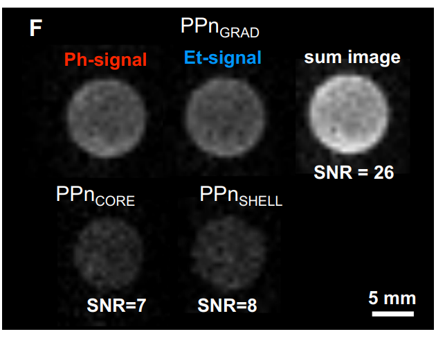
Figure 2. 31 P MRI of PPnGRAD micelles (top row). The signals of both Ph- and Et-units were added to a sum image, resulting in the highest SNR. The images of block copolymer micelles PPnCORE and PPnSHELL are shown at the same intensity scale for comparison. Polymer content 4 wt% ( 31 P 0.1 M in PPnCORE, 2×0.1 M in each block of PPnGRAD; and 0.05 M 31 P In PPnSHELL; similar polymer concentration was chosen. FOV 20 × 20 mm2 , matrix 64 × 64, 9.4 T.
Novel Aspects:
The use of mCSSI for 31P imaging represents a significant advancement over traditional imaging techniques, allowing for simultaneous detection of multiple signals without interference, thus enhancing the diagnostic capability of the micelles.
Experiment 3: In Vivo Imaging and Biodegradation Studies
Key Steps:
- Animal Model Selection: Manduca sexta caterpillars were chosen as the model organism for in vivo experiments due to their comparable hemolymph volume to small mammals.
- Injection of Micelles: The micelles were injected into the dorsal vessel of the caterpillars, allowing for real-time monitoring of their distribution.
- MRI Scanning: Post-injection, 31P mCSSI MRI scans were performed at specified time points to observe the circulation and degradation of the micelles.
Data Collection and Analysis:
MRI data were collected at intervals of 0 and 24 hours post-injection, and the presence of the micelles in hemolymph was confirmed through overlaying anatomical 1H MRI images with 31P images.
Result:
The micelles circulated in the hemolymph for 24 hours, demonstrating their stability and stealth properties. Additionally, degradation products were identified in fecal samples via 31P NMR spectroscopy, confirming in vivo biodegradation.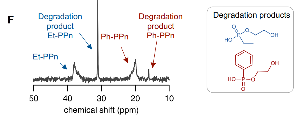
Figure 3. 31P NMR spectrum of feces (D2O, 158 MHz) collected after 24 h shows the characteristic signals of degradation products confirming that PPnGRAD agents are biodegradable. Color scale in a.u.
Novel Aspects:
This experiment highlighted the biodegradability of the polyphosphoester micelles in a living organism, a critical advantage over traditional drug delivery systems that often accumulate in organs and pose toxicity risks.
Experiment 4: Drug Delivery Efficacy
Key Steps:
- Loading of Therapeutic Agent: The hydrophobic drug PROTAC was encapsulated in the synthesized micelles using a nanoprecipitation technique.
- Stability Testing: The stability of the drug-loaded micelles was assessed over two months through DLS and visual inspection.
- Cell Viability Assay: The efficacy of the drug-loaded micelles was evaluated using HeLa cancer cells to determine their impact on cell viability and apoptosis induction.
Data Collection and Analysis:
Viability was measured using standard assays, and statistical analysis was performed to compare the effects of free PROTAC and PROTAC-loaded micelles on cell viability and apoptosis rates.
Result:
The drug-loaded micelles maintained stability over two months and exhibited comparable cytotoxicity to free PROTAC, reducing the viability of HeLa cells significantly.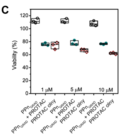
Figure 4. Viability of cancer cells (HeLa) was reduced upon the administration of PROTAC-loaded micelles to a comparable level as with free PROTAC. Unloaded PPnGRAD micelles were used as a control. The viability values are normalized to untreated controls. n = 3 (data presented as mean ± SD). D Flow cytometry shows that cancer cells treated with PROTAC-loaded micelles (1 µM ARV825) start entering an early apoptosis phase
Novel Aspects:
This experiment established the micelles as effective drug delivery systems while simultaneously serving as imaging agents, demonstrating their potential for theranostic applications—a significant improvement over conventional delivery systems that often lack imaging capabilities.
Overall, the methodology employed in this study effectively combined polymer synthesis, advanced imaging techniques, and drug delivery, ensuring replicability and robustness in the findings.
Conclusion
The research presents groundbreaking findings regarding the use of biodegradable polyphosphoester micelles as dual-function agents for background-free MRI and drug delivery. The micelles not only provide a solution to the environmental issues associated with traditional imaging agents but also enhance the possibilities for targeted drug therapy.
While the study lays a strong foundation for future applications, it acknowledges limitations such as the need for further testing in mammalian models to fully understand the biodistribution and degradation of the micelles. Future research could explore the scalability of these micelles for clinical applications, as well as their potential use in other therapeutic contexts, broadening the impact of this innovative approach in biomedicine.
In summary, the findings of this research pave the way for the development of sustainable, effective, and multifunctional materials in the fields of imaging and drug delivery.
Reference
Koshkina, Olga, et al. “Biodegradable polyphosphoester micelles act as both background-free 31P magnetic resonance imaging agents and drug nanocarriers.” Nature communications 14.1 (2023): 4351