Editor: Nina
Key Preview
Research Question:
- Can cerium oxide nanoparticles (CeO2NPs) serve as a safe and effective alternative to iodine-based contrast agents for X-ray CT imaging? Specifically, do these nanoparticles provide improved imaging contrast with reduced toxicity and prolonged residence times in vivo?
Research Design and Strategy:
- Utilized in vivo models with athymic nude mice bearing xenografted sarcoma tumors.
- Conducted comparative analyses of CeO2NPs and conventional iodine-based contrast agents to assess imaging efficiency and safety.
- Investigated biodistribution of nanoparticles across organs to evaluate retention and clearance.
Method:
- Synthesized albumin-stabilized CeO2NPs (~5 nm) to improve biocompatibility and prevent aggregation.
- Administered nanoparticles via two routes: intravenous (IV) and intratumoral (IT).
- Used X-ray CT imaging for real-time assessment of imaging contrast and ICP-MS for organ biodistribution quantification.
Key Results:
- CeO2NPs demonstrated up to 10-fold improved contrast compared to iodine-based agents at much lower concentrations.
- After IV administration, 85% of the dose accumulated in the liver and spleen after seven days. For IT administration, 99% of the dose remained in the tumor for the same period.
- CeO2NPs showed superior solubility, dispersion, and non-immunogenicity compared to iodine-based agents, which cleared rapidly through renal excretion.
Significance of the Research:
- Demonstrated the feasibility of CeO2NPs as safe contrast agents for prolonged, high-quality imaging.
- Highlighted their radioprotective and antioxidant properties, which reduce the risk of radiation-induced damage.
- Suggested potential applications for CeO2NPs in pediatric and other sensitive imaging scenarios, as well as for studying tumor growth dynamics.
Introduction
The study builds on two decades of research aimed at developing alternative contrast agents that address the limitations of iodine-based compounds, which are associated with rapid clearance from the bloodstream and potential nephrotoxicity. The evolution of imaging technology necessitates advancements in contrast agents to improve diagnostic capabilities while ensuring patient safety. Cerium oxide nanoparticles, with their unique catalytic properties, emerge as a promising candidate for this purpose.
Research Team and Objective

The research team, led by Ana García and comprising experts from institutions such as Vall d’Hebron Hospital Universitari and the Institut Català de Nanociència i Nanotecnologia, aimed to explore the potential of CeO2NPs in enhancing X-ray imaging capabilities. Their objective was to evaluate not only the imaging efficacy but also the safety and biodistribution profile of these nanoparticles in a preclinical setting.
Experimental Process
· Synthesis of Albumin-Stabilized CeO2NPs:
- Procedure: CeO2NPs (~5 nm) were chemically synthesized by precipitating cerium nitrate in a basic solution and conjugated with murine serum albumin (MSA) to improve solubility and biocompatibility.
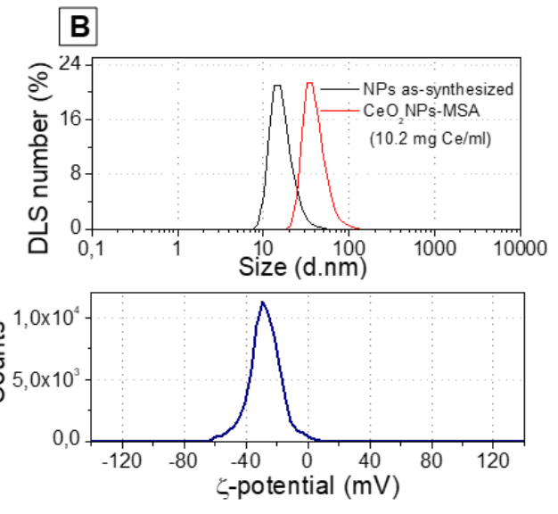
- Result: Achieved stable colloidal nanoparticles with a hydrodynamic diameter of 38.7 ± 1.9 nm, a ζ-potential of −28.2 mV, and no aggregation.

Figure 1. Top: Dynamic light scattering (DLS) measurements showing the size distribution profile of CeO2NPs by Number fits. As-synthesized CeO2NPs are small aggregates with a number mean of 17.6 nm; once conjugated with albumin and concentrated (10.2mg Ce/ml), the number mean increases up to 38.7 nm. Bottom: ζ-potential profile (-28.2 ± 0.73 mV; media conductivity 0.74 ± 0.02 mS/cm; pH 7.4). Data provided is the mean of three independent measurements.
- Significance: Stability and biocompatibility are crucial for safe in vivo applications.
- Innovation: Enhanced catalytic activity by optimizing nanoparticle size.
· Intravenous Administration of CeO2NPs:
- Procedure: 200 µL of CeO2NPs-MSA (10 mg Ce/mL) was injected into the tail veins of nude mice bearing sarcoma tumors. Imaging was performed at multiple time points (15 min, 1 h, 24 h, 7 days).
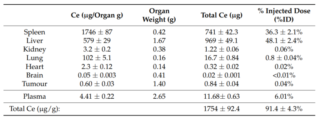
- Result: Liver and spleen exhibited significant nanoparticle retention (85% of the injected dose recovered after 7 days). In contrast, iodine-based agents were rapidly cleared through the kidneys.

Table 1. ICP-MS analysis of Ce mass in mice organs (7 days post-i.v. injection), the weight of organs, total Ce mass in each organ and the corresponding %ID(2040±98mg Ce)
- Significance: The prolonged residence of CeO2NPs in target tissues enables extended imaging windows, reducing the need for repeat dosing.
- Innovation: Non-immunogenic and non-toxic profile with superior retention compared to conventional agents.
· Intratumoral Administration of CeO2NPs:
- Procedure: 70 µL of CeO2NPs-MSA was injected directly into the tumors. Imaging and ICP-MS were conducted over seven days to monitor biodistribution and contrast enhancement.
- Result: A 10-fold increase in contrast was observed (from 75 HU to 747 HU) within 15 minutes of injection, persisting for seven days. Biodistribution analysis confirmed 99% retention of Ce in the tumor.

Table 2. CT contrast analysis (HU and volume) of the tumoral region after intratumoral injection of CeO2NPs-MSA solution (70 µL, 10 mg Ce/mL).
- Significance: Persistent tumor retention allows for detailed monitoring of structural tumor changes over time.
- Innovation: Enabled differentiation between tumor progression and pseudoprogression.
· CT Imaging:
- Procedure: CT scans were performed using a Quantum FX micro-CT system. Serial coronal and axial views were analyzed for regions of interest (liver, spleen, kidney, tumor, etc.).
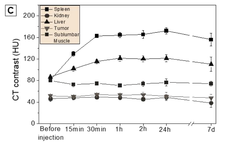
- Result: CeO2NPs provided significantly higher contrast density compared to iodine agents, especially in liver and spleen regions. Tumor imaging revealed sustained high contrast over time, with minimal spread to adjacent tissues.

Figure 2. Temporal evolution of CT contrast density values (HU) of the tissues under study
- Significance: High contrast density facilitates precise imaging of disease dynamics, including tumor progression.
- Innovation: First demonstration of CeO2NPs achieving long-lasting contrast enhancement with low toxicity.
· Comparison with Iodine-Based Agents:
- Procedure: Iopamidol®-370 was used as a control contrast agent. Imaging was conducted to compare biodistribution and contrast levels.
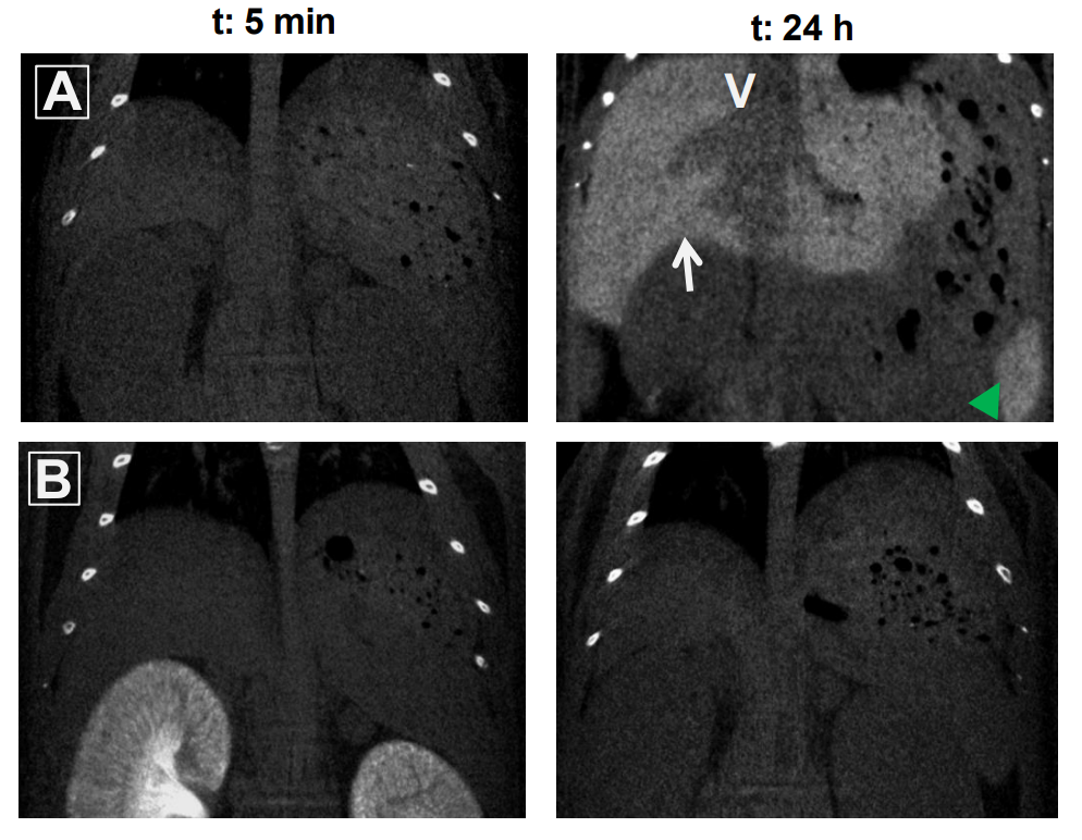
- Result: Iodine agents rapidly cleared from kidneys within 24 hours, whereas CeO2NPs persisted in liver, spleen, and tumor for seven days.

Figure 3. CT coronal images 5 min and 24 h after intravenous injection of CeO2NPs-MSA (panel A) and the commercial iodine contrast agent Iopamidol®-370 (150 µl of 175 µg I/ml) (panel B). CeO2NPs accumulate progressively on the liver (white arrow) and spleen (green arrowhead) throughout 24 h after injection, when it can be clearly distinguished the vena cava (V) and the intrahepatic vessels. On the contrary, the iodine contrast is already accumulated in kidneys 5 min post-injection and completely absent 24 h later.
- Significance: Highlights the limitations of iodine-based agents, such as short imaging windows and potential nephrotoxicity.
- Innovation: Demonstrated the superiority of CeO2NPs in terms of retention, contrast, and safety profile.
· Tumor Growth Monitoring:
- Procedure: Tumor growth was tracked using CT scans and quantitative analysis. Contrast intensity and tumor volume were measured over seven days.
- Result: Tumor volume increased from 700 mm³ to 1,500 mm³, with CeO2NPs enabling detailed visualization of structural changes within the tumor.
- Significance: Enables non-invasive, longitudinal monitoring of tumor dynamics.
- Innovation: Establishes CeO2NPs as a diagnostic tool for analyzing tumor microenvironment and growth.
Conclusion
The findings of this study highlight the potential of CeO2NPs as a revolutionary contrast agent for X-ray CT imaging. Their high contrast enhancement, low toxicity, and prolonged residence time within target tissues position them as a viable alternative to traditional iodine-based agents. However, further research is needed to fully understand their long-term effects and optimize their use in clinical settings. The study opens avenues for future investigations into the clinical applications of CeO2NPs, particularly in improving imaging outcomes while safeguarding patient health.
Reference
García, Ana, et al. “Nanoceria as Safe Contrast Agents for X-ray CT Imaging.” Nanomaterials 13.15 (2023): 2208.
